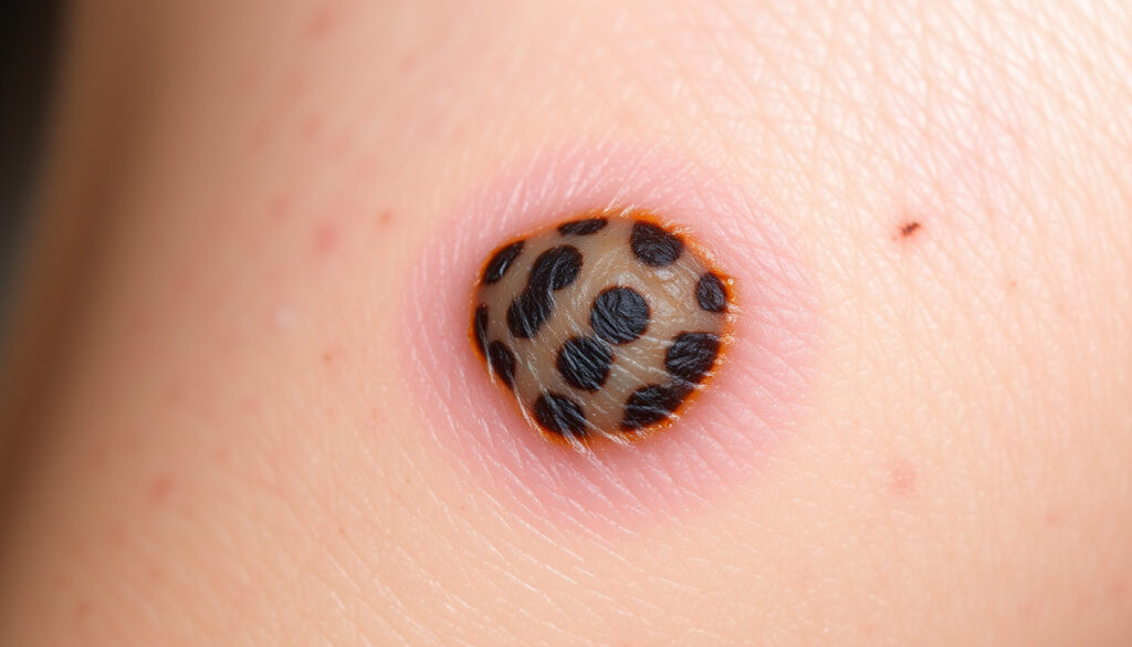
What Does Melanoma Look Like – Visual Guide to Early Detection
Melanoma represents the most serious form of skin cancer, yet when identified early, it carries a survival rate exceeding 99 percent. Recognizing what melanoma looks like can quite literally save lives, making visual literacy around skin changes a critical health skill for everyone.
Unlike more common skin cancers that develop slowly in one location, melanoma can arise anywhere on the body and spreads more rapidly if left undetected. The challenge lies in its variability—melanoma does not always present as the stereotypical dark mole. Understanding its many appearances empowers individuals to seek medical evaluation promptly when something seems amiss.
This guide walks through the visual characteristics, warning signs, and specific presentations of melanoma across different skin types and body locations, grounded in guidance from dermatological authorities and cancer organizations.
What Does Melanoma Look Like? Key Visual Characteristics
Melanoma lesions frequently display identifiable visual features that distinguish them from benign moles. While no single characteristic guarantees a melanoma diagnosis, several patterns warrant particularly close attention and professional evaluation.
Asymmetry
One half of the lesion does not match the other when divided by an imaginary line through the center.
Border Irregularity
Edges appear blurred, ragged, notched, or scalloped rather than smooth and well-defined.
Color Variation
Multiple shades within the same lesion—brown, tan, black, red, white, or blue—rather than uniform coloring.
Evolution
Changes in size, shape, color, elevation, or new symptoms like bleeding, itching, or crusting over weeks to months.
Not all melanomas follow this pattern perfectly. Certain aggressive subtypes may lack pigment entirely or grow downward into deeper skin layers before showing surface changes. According to the Skin Cancer Foundation, the presence of any one of these characteristics—particularly evolution—should prompt a dermatologist visit.
- Early detection through self-examination improves cure rates significantly, with five-year survival exceeding 99 percent for thin, localized melanomas.
- Melanoma can develop in sun-exposed areas as well as regions typically protected from ultraviolet radiation, including the scalp, groin, and soles of the feet.
- Approximately 20 to 30 percent of melanomas are amelanotic, meaning they lack dark pigment and may appear pink, red, or skin-colored.
- The “ugly duckling” concept identifies spots that look notably different from all other moles on an individual’s body, regardless of how they measure against standard criteria.
- Regular skin self-examinations complement but do not replace professional dermatological evaluations.
- Melanoma on darker skin tones may present differently, often appearing as dark brown, black, or gray spots with irregular borders rather than the classic pigmented lesion.
| Characteristic | Details |
|---|---|
| Typical Diameter | Greater than 6 millimeters (roughly the size of a pencil eraser), though earlier-stage lesions may be smaller |
| Color Possibilities | Brown, tan, black, red, white, blue, or multiple shades within a single lesion |
| Common Locations in Men | Trunk (back and chest), head, neck |
| Common Locations in Women | Legs, arms |
| Stage 1 Thickness | Less than 1 millimeter, confined to the epidermis |
| Amelanotic Prevalence | 20-30% of all melanoma cases |
| Nodular Misdiagnosis Rate | 25-89% initially mistaken for benign conditions |
| Growth Pattern | Superficial spreading (horizontal) or vertical (nodular), the latter being more aggressive |
What Is the ABCDE Rule for Spotting Melanoma?
The ABCDE rule provides a simple framework for evaluating moles and skin lesions at home. Developed by dermatologists and widely promoted by cancer organizations, these five criteria help individuals identify characteristics that merit professional assessment.
Breaking Down Each Criterion
Asymmetry becomes evident when one half of a lesion fails to mirror the other half. Drawing an imaginary line through the center of the spot should produce matching halves if the lesion is symmetrical. A melanoma typically displays lopsided or uneven halves, as noted by the MyMelanomaTeam resources.
Border irregularity manifests as edges that are blurred, ragged, notched, or scalloped rather than smooth and well-defined. Benign moles usually maintain consistent, clean borders throughout their perimeter. The transition zone between a melanoma and surrounding skin often lacks the clear demarcation seen in harmless growths.
Color variation within a single lesion serves as another warning sign. While harmless moles typically display uniform brown coloring throughout, melanomas frequently contain multiple shades or colors. These may include different shades of brown, tan, black, red, white, or blue appearing within the same spot. According to the Skin Cancer Foundation, this uneven pigmentation distribution distinguishes concerning lesions from common moles.
Diameter greater than 6 millimeters—approximately the width of a pencil eraser—raises suspicion, though melanomas can certainly present smaller, particularly in their earliest stages. Lesions darker than surrounding spots also warrant attention regardless of size.
Evolution represents perhaps the most critical criterion. Any spot that changes in size, shape, color, elevation, or texture over weeks or months demands evaluation. New symptoms such as itching, bleeding, oozing, or crusting also indicate potential concern. The concept of the “ugly duckling” lesion—any spot that looks notably different from all others on an individual’s body—supplements the ABCDE framework for identification.
The ABCDE rule applies most reliably to superficial spreading melanoma, the most common type. Nodular and amelanotic melanomas may not conform to these visual criteria, making regular dermatological examinations especially valuable for early detection of these aggressive subtypes.
Distinguishing Melanoma from Benign Moles
Benign moles typically exhibit symmetry, smooth borders, uniform color, and remain stable over time without exceeding 6 millimeters in diameter. They often appear during childhood or early adulthood and may fade with age. In contrast, melanomas frequently emerge as new growths in adulthood or represent changes in existing moles. The Cancer Treatment Centers of America emphasizes that any new, changing, or unusual skin growth warrants professional evaluation regardless of how it measures against these general characteristics.
What Does Early Stage Melanoma Look Like?
Stage 1 melanoma represents the earliest invasive phase of this cancer, characterized by thin lesions that have not yet penetrated deeply into the skin. Understanding their appearance proves essential for catching the disease when treatment proves most effective.
Stage 1 Melanoma Characteristics
Stage 1 melanomas measure less than 1 millimeter in thickness and show no ulceration of the outer skin layer. They remain confined to the epidermis—the outermost layer of skin—with no spread to lymph nodes or distant organs. This early-stage presentation carries an excellent prognosis when identified and treated promptly.
Visually, stage 1 melanomas often resemble early-stage superficial spreading melanoma, displaying the asymmetry, irregular borders, and color variations described by the ABCDE rule. The lesion may have recently emerged or undergone subtle changes in an existing mole. Size at this stage can vary, though many measure under 6 millimeters. Some stage 1 melanomas may present as pigmented patches with slightly raised or textured areas, while others manifest as small, dark spots with concerning border characteristics.
It is worth noting that certain subtypes of early melanoma—including amelanotic and nodular variants—may not display classic pigmentation patterns. These lesions can appear as pink, red, or skin-colored bumps or flat spots, making them easier to dismiss as harmless. The Instalab medical resources document how amelanotic melanoma frequently presents as a pink or flesh-colored lesion, potentially with subtle gray or brown edges that become more apparent upon closer examination.
When caught at stage 1, melanoma five-year survival rates exceed 99 percent. Regular skin self-examinations, particularly for individuals with risk factors such as fair skin, numerous moles, or significant sun exposure history, significantly increase the likelihood of identifying these early-stage lesions before progression occurs.
Where Does Melanoma Typically Appear and Variations?
Melanoma can develop anywhere on the body, though certain locations show higher prevalence depending on sex and skin type. Understanding these patterns helps focus examination efforts on higher-risk areas while remaining vigilant across all skin surfaces.
Common Sites by Gender
Research documented by the MyMelanomaTeam reveals distinct patterns in melanoma location between sexes. In men, melanomas most frequently appear on the trunk—the back and chest—followed by the head and neck region. Women tend to develop melanomas more commonly on the legs and arms. These patterns likely reflect differences in sun exposure habits and clothing choices rather than inherent biological differences.
Beyond these typical locations, melanoma can occur in areas rarely exposed to sunlight: the scalp, ears, genitals, palms of the hands, and soles of the feet. This variability underscores why comprehensive skin examinations—including areas not routinely visible—prove essential for early detection.
Melanoma Appearance on Darker Skin Tones
Melanoma on individuals with black or brown skin may present distinctly from classic descriptions. A study published in the National Institutes of Health database documents that melanomas on darker skin tones often appear as dark brown, black, or gray spots with irregular borders, potentially mimicking benign moles more closely than lighter skin presentations.
Acral lentiginous melanoma (ALM) represents a subtype particularly relevant to individuals with darker skin. This cancer commonly develops on the palms, soles, and under nails—areas where sun exposure plays no role in development. Early ALM typically presents as a flat, discolored patch that gradually becomes more irregular and may thicken over time. Any new or changing spot on these locations merits dermatological evaluation, particularly when asymmetry or color variation develops.
Melanoma under nails (subungual melanoma), on the soles, and in genital regions often escapes early detection due to less routine examination. Any pigmented streak under a nail, new dark spot on the sole, or unusual lesion in hidden areas should prompt professional assessment regardless of how it conforms to standard visual criteria.
Common Melanoma Types and Their Appearances
Melanoma encompasses several distinct subtypes, each with characteristic visual features and growth patterns. Recognizing these variations improves detection across the full spectrum of this disease.
Superficial Spreading Melanoma
The most common form, superficial spreading melanoma, typically follows the classic ABCDE pattern most closely. These lesions usually begin as flat or slightly raised pigmented patches that expand horizontally across the skin surface. Color variation commonly develops over time, with shades of brown, black, red, white, and blue appearing within the same lesion. Border irregularity becomes increasingly apparent as the lesion grows. This type most frequently appears on the legs in women and the trunk in men, though it can develop anywhere.
Nodular Melanoma
Nodular melanoma represents a more aggressive variant characterized by rapid vertical growth into deeper skin layers. Visually, these lesions typically present as firm, raised bumps or nodules rather than flat patches. According to documented cases on DermNet NZ, nodular melanomas are frequently amelanotic, appearing pink, red, or flesh-colored rather than heavily pigmented. This lack of classic coloring, combined with their raised appearance, often leads to initial misdiagnosis—studies indicate misdiagnosis rates between 25 and 89 percent when these lesions first present.
Common locations include the scalp, thighs, and shoulders. The rapid growth pattern means that nodular melanoma may double in size within months, underscoring the importance of prompt evaluation for any new or rapidly changing raised lesion.
Amelanotic Melanoma
Amelanotic melanoma comprises approximately 20 to 30 percent of all cases, distinguished by its lack of pigment production. These lesions may appear pink, red, skin-colored, or have subtle gray-brown edges, as documented by Cancer Treatment Centers of America. The absence of dark coloring means these melanomas frequently evade the ABCDE framework, which relies heavily on color variation as a warning sign.
Dermoscopy—a specialized skin examination technique—may reveal polymorphous vessels, structureless white or pink areas, and occasional brown dots in amelanotic lesions. These findings differ markedly from the pigment networks visible in typical melanomas. Under clinical observation, amelanotic lesions may display linear-irregular, arborizing, or looped vessels that reflect their aggressive nature.
Lentigo Maligna and Other Variants
Lentigo maligna melanoma typically develops on sun-damaged skin, particularly the face and neck of older adults. This slow-growing variant begins as a flat, tan or brown patch with irregular borders that may resemble a large freckle. Over years, the lesion may develop darker pigmentation and raised areas, signaling progression to invasive melanoma. Due to its slow horizontal growth pattern, lentigo maligna may be present for years before becoming concerning.
Acral lentiginous melanoma, discussed previously in relation to darker skin tones, presents as a flat, discolored area that gradually becomes more irregular and may thicken. This subtype appears on palms, soles, and beneath nails regardless of sun exposure history.
No two melanomas appear identical, and significant variation exists between subtypes and individuals. The presence of any new, changing, or unusual skin lesion warrants professional evaluation. Dermatologists use specialized tools including dermoscopy and, when necessary, biopsy to confirm diagnoses that visual examination alone cannot establish.
How Does Melanoma Progress Over Time Without Detection?
Understanding melanoma progression helps contextualize why early identification matters. Without intervention, melanoma advances through distinct stages, with each stage representing greater thickness, depth of invasion, and spread.
- Stage 0 (In Situ): Cancer cells remain confined to the epidermis, the outermost skin layer, without invasion into deeper tissues. At this stage, melanoma appears as an abnormal patch or spot with concerning visual features but has not yet acquired the ability to spread.
- Stage 1: The lesion has invaded the dermis, the layer beneath the epidermis, but remains thin (less than 1 millimeter) with no ulceration. Survival rates at this stage exceed 99 percent with appropriate treatment, typically surgical removal with clear margins.
- Stage 2: The melanoma has grown thicker (between 1 and 4 millimeters) or shows ulceration, indicating more aggressive behavior. While still localized, these characteristics increase recurrence risk. Additional treatments such as sentinel lymph node biopsy may be recommended.
- Stage 3: Cancer has spread to nearby lymph nodes or lymph vessels near the original site. Treatment expands to include lymph node removal and potentially adjuvant therapies such as immunotherapy to reduce recurrence risk.
- Stage 4: Melanoma has metastasized to distant organs—lungs, liver, brain, or other sites—making cure significantly more challenging. Modern immunotherapy and targeted therapies have improved outcomes, but stage 4 disease requires ongoing management rather than curative treatment.
The timeline for progression varies considerably between subtypes. Nodular melanomas grow rapidly, potentially advancing through stages within months, while superficial spreading and lentigo maligna types may take years to progress. Amelanotic melanoma often evades early detection due to its subtle appearance, frequently presenting at more advanced stages than pigmented variants.
What Can Be Confidently Identified Versus What Requires Professional Evaluation?
Clear communication about what individuals can reasonably assess at home versus what demands medical expertise helps set appropriate expectations and promotes timely professional consultation.
| Established Visual Indicators | Features Requiring Professional Assessment |
|---|---|
| Obvious asymmetry where one half differs markedly from the other | Subtle pigment variations that may or may not represent melanoma |
| Clearly irregular, ragged, or scalloped borders | Dermoscopic patterns visible only with specialized equipment |
| Multiple distinct colors within a single lesion | Lesions without classic ABCDE features but persisting or changing |
| Size exceeding 6 millimeters with other concerning features | Pigment patterns in nail beds requiring Wood’s lamp examination |
| Documented change in size, shape, or color over weeks to months | Cellular characteristics determinable only through biopsy pathology |
| New symptoms such as bleeding, itching, or crusting in a skin lesion | Depth of invasion, which guides staging and treatment decisions |
Self-examination serves as a valuable screening tool but cannot replace professional dermatological evaluation. Dermatologists possess training to identify subtle warning signs invisible to untrained observers, access to dermoscopy that reveals patterns beneath the skin surface, and the ability to perform biopsies that provide definitive diagnosis. The Centers for Disease Control and Prevention recommends regular skin examinations as part of comprehensive skin cancer prevention, while acknowledging that professional assessment remains essential for any concerning findings.
Understanding Melanoma Risk Factors and Prevention Context
While visual identification focuses on lesion characteristics, understanding risk factors provides important context for who should maintain heightened vigilance. Melanoma develops through a complex interaction of genetic predisposition and environmental exposure.
Ultraviolet radiation from sunlight and tanning beds represents the primary environmental risk factor for most melanoma types. Intermittent, intense sun exposure—particularly during childhood and adolescence—increases risk more significantly than gradual chronic exposure. Geographic location, altitude, and reflective surfaces amplify UV intensity and consequent risk.
Individual characteristics influence susceptibility considerably. Fair skin that burns easily, blue or green eyes, blonde or red hair, numerous freckles, and high mole counts all correlate with increased melanoma risk. A family history of melanoma or previous melanoma diagnosis substantially elevates personal risk. Immunosuppression, whether from medication or medical conditions, also increases vulnerability to skin cancer development.
Prevention strategies complement visual monitoring. Daily broad-spectrum sunscreen application, protective clothing including wide-brimmed hats, seeking shade during peak sun hours, and avoiding tanning beds all reduce melanoma risk. Monthly skin self-examinations allow individuals to track their moles and identify changes promptly. Annual professional skin examinations provide expert evaluation, particularly important for those with elevated risk profiles.
What Do Medical Authorities Recommend for Melanoma Screening?
Major medical organizations provide consistent guidance regarding melanoma detection approaches, emphasizing both self-examination and professional evaluation as complementary components of effective screening.
“The earlier melanoma is found, the easier it is to treat. Learn the warning signs of melanoma so you can see a doctor right away if you notice any changes to your skin.”
— Centers for Disease Control and Prevention
“Melanoma can appear on skin that hasn’t been exposed to the sun. Examine your entire body and don’t forget to check your scalp, palms, fingernails, toenails, and the soles of your feet.”
— American Academy of Dermatology
The Mayo Clinic emphasizes that melanoma can develop anywhere on the body, including areas with minimal sun exposure, and recommends whole-body skin examinations as part of routine preventive care. They note that individuals with risk factors should discuss the appropriate frequency of professional skin examinations with their healthcare providers.
For those seeking to understand how this topic relates to broader health questions, exploring What Does Pepto Bismol Do may provide additional perspective on how different health conditions present and require professional evaluation for accurate diagnosis.
Key Takeaways for Identifying Melanoma
Recognizing melanoma begins with understanding that this cancer takes many forms beyond the stereotypical dark mole. The ABCDE framework—asymmetry, border irregularity, color variation, diameter exceeding 6 millimeters, and evolution—provides a useful starting point for self-examination, though awareness of its limitations proves equally important. Aggressive subtypes like nodular and amelanotic melanoma frequently deviate from these classic criteria, presenting instead as pink, red, or flesh-colored raised bumps that may grow rapidly.
Melanoma can develop anywhere on the body, with typical locations varying by sex and skin type. Higher-risk areas include the trunk and head in men, the legs in women, and the palms, soles, and nail beds regardless of sun exposure history. Individuals with darker skin tones should pay particular attention to acral areas, where melanoma may appear differently than in lighter-skinned populations.
Self-examination serves as a valuable first-line screening tool, but any new, changing, or unusual skin lesion warrants professional dermatological evaluation. Dermatologists possess specialized training and equipment—including dermoscopy and biopsy capability—that enable accurate diagnosis beyond what visual inspection alone can achieve. Regular professional examinations complement monthly self-checks, particularly for individuals with elevated risk profiles.
Early-stage melanoma carries survival rates exceeding 99 percent, making prompt identification one of the most powerful tools available in fighting this disease. Knowledge of what melanoma looks like across its many presentations, combined with vigilant monitoring and timely professional consultation, represents the most effective strategy for early detection and successful treatment outcomes.
Frequently Asked Questions
Is melanoma raised or flat?
Melanoma presentation varies by subtype. Superficial spreading melanoma often begins flat and may become raised over time, while nodular melanoma typically presents as a firm, raised bump from the outset. Early-stage lesions may be flat or slightly elevated.
Are melanoma spots itchy?
Itching can occur with melanoma, particularly in more advanced lesions. New symptoms such as itching, bleeding, oozing, or crusting of any skin lesion warrant professional evaluation, though itching alone does not confirm melanoma.
What color is melanoma?
Melanoma may display multiple colors—brown, black, tan, red, white, or blue—or may lack pigment entirely. Amelanotic melanoma appears pink, red, or skin-colored. The presence of multiple shades within a single lesion represents a warning sign.
Does melanoma always bleed?
Bleeding is not always present, particularly in early-stage melanoma. However, any lesion that bleeds, oozes, crusts, or develops these symptoms warrants professional assessment. Advanced lesions are more likely to exhibit these characteristics.
Can melanoma be pink?
Yes. Amelanotic melanoma—comprising 20 to 30 percent of cases—lacks dark pigment and typically appears pink, red, or flesh-colored. These lesions may have subtle gray or brown edges and frequently evade the ABCDE criteria.
How fast does melanoma grow?
Growth rates vary significantly by subtype. Nodular melanoma can grow rapidly over weeks to months, while superficial spreading melanoma typically progresses more slowly over months to years. Any rapidly changing lesion demands urgent evaluation.
Does melanoma look like a pimple?
Nodular and amelanotic melanoma can resemble bumps or pimple-like lesions, which contributes to their frequent misdiagnosis. A spot that persists beyond several weeks, grows rapidly, or differs from typical acne should be evaluated by a dermatologist.
Where does melanoma usually start?
Melanoma can originate anywhere on the body, including areas without sun exposure. Most commonly, it develops in sun-exposed areas: on men’s trunks, women’s legs, and on the face and arms. It can also begin in the eyes, under nails, and on soles and palms.
More related posts
 When Does Gen Z End? Birth Years & Age Range
 J.J. Spaun Biography: Ethnicity, Wife, Age, Net Worth & Career
 Open Farm Dog Food: Vet Review & Top Comparisons
 Red Dresses for Women: Styles, Lengths & Irish Shops
 What Does Pepto Bismol Do – Uses, How It Works, Side Effects
 Oat Milk vs Almond Milk: Which Is Healthier? Full Comparison
 Sonic the Hedgehog 2020: Box Office, Cast, Where to Watch
 Amazon Customer Service Number – US, UK & Ireland Info
